MELANOMA: Early diagnosis and treatment crucial
Treatment will begin and end in a dermatologist’s office for most patients who get melanoma.
They won’t need the array of advanced treatments discussed in an accompanying article. They won’t see medical or surgical oncologists.
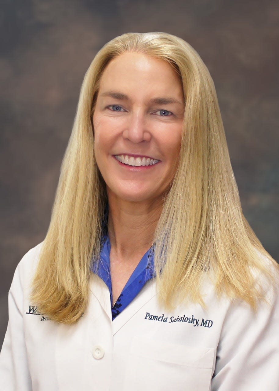
“People get scared of that word, melanoma, but if it’s caught in the early stages, it’s curable,” said Dr. Pamela Sakalosky, a Watson Clinic dermatologist.
Highly curable, in fact.
The five-year survival rate for localized melanoma that hasn’t spread beyond the skin where it was found is as high as 99 percent.
This is a sharp contrast to the 30 percent for people whose cancer has spread to distant parts of the body (lungs, liver or skin in other areas). The rate is 68 percent if cancer is found when it’s spread to nearby areas or lymph nodes.
Early diagnosis and treatment make a difference. That’s why it’s extremely important for people to monitor changes on their skin, see a dermatologist about suspicious ones and not let fear cause them to delay.
“The earlier we get these cancers, the more chance there is to be cured,” said Dr. Manuel Molina-Vega, surgical oncologist at Lakeland Regional Health Hollis Cancer Center.

Primary care doctors also need to look at their patients’ skin and identify any unusual moles or changes, Sakalosky said.
“The color of the area is usually pigmented, oftentimes showing different shades of brown with variations in color,” she said when asked about differentiating melanoma from basal or squamous cell skin cancers.
The latter two usually are red and more raised, she said, although squamous also can look like an age spot or a wart.
All should be followed up, whether they are melanoma (the most deadly) or the more common basal or squamous cell. Better to go in with one that turns out to be a benign condition than ignore the one that may be cancer.
The often reported A,B, C, D, E chart is helpful in detecting melanoma:
• Asymmetry, when half the spot doesn’t look like the other half
• Border, as in an irregular, scalloped or poorly defined
• Color varies from one area to the next, such as shades of tan, brown or black.
• Diameter of a melanoma usually is about the size of a pencil eraser at diagnosis
• E is for evolving. The spot looks different or is changing in size, shape or color.
Age-adjusted rates for new melanoma of the skin cases rose 1.2 percent a year, on average, over 2010-2019, according to the National Cancer Institute.
The good news is that age-adjusted death rates were falling, too.
“Melanoma mortality dropped steeply from 2015 to 2019, by about 4 percent a year, because of advances in treatment,” the American Cancer Society says in its 2022 report.
Area oncologists interviewed confirm they are seeing more cases of melanoma, as is Sakalosky.
“I am definitely seeing more melanoma, thankfully more early stage melanoma,” Sakalosky said.

Dr. Shalini Mulaparthi, medical oncologist at Watson Clinic Cancer and Research Center, also sees an increase, The level of severity is higher for cases she and other medical or surgical oncologists treat.
“At our cancer center, during the past five years, we have seen a rising number of advanced melanoma cases that we’ve been treating,” Mulaparthi said.
She and Molina-Vega also are supportive of skin self-examination and regular skin exams by a dermatologist .
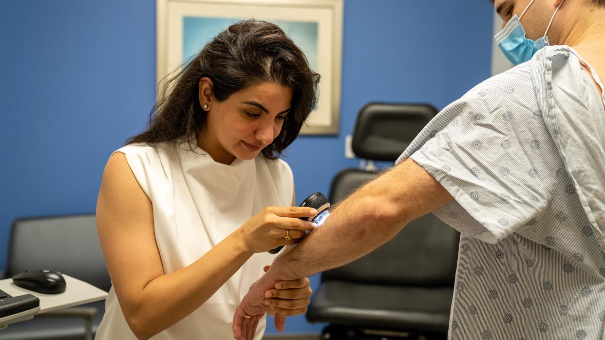
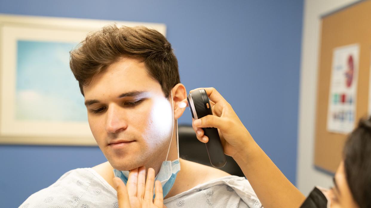
The first stop for melanoma diagnosis and treatment is a dermatologist’s office for a skin exam, removal of skin from suspicious patches and sending those samples for a biopsy.
The melanoma skin cancer is measured to get its depth, called a Breslow thickness, Sakalosky said. That’s from the surface of the skin to the deepest point of the tumor.
For most melanomas, if the depth is less than 0.7 millimeters, she said, the patient doesn’t need to be referred for a sentinel lymph node biopsy. If it’s deeper, she refers to a surgeon for a sentinel lymph node biopsy to make sure the melanoma hasn’t spread.
Sentinel lymph nodes are the ones closest to the cancer.
For thicker melanomas and those with a positive sentinel lymph node, the patient is referred to oncology.
Thicker melanomas have a greater chance of spreading.
With early stage melanomas, surgery to remove the cancer and a margin of normal skin around it is the typical treatment. Width depends on the thickness and location of the melanoma.
These excision surgeries of melanoma that hasn’t spread beyond the skin can and usually are done by dermatologists. Patients stay awake.
What if cancer cells are found in the sentinel lymph nodes?
It used to be common to remove all lymph nodes to which intermediate thickness melanoma had spread.
MSLT-II, an earlier trial in which Lakeland Regional took part, significantly changed that. The trial determined doing that wasn’t leading to increased survival, so the practice was stopped, Molina-Vega said.
Cancer-positive lymph nodes now left inside after surgery are monitored with ultrasound instead. Therapies such as immunotherapy also may be used.
Robin Williams Adams, robinwadams99@yahoo.com
This article originally appeared on The Ledger: Early diagnosis and treatment crucial for melanoma patients
